Knee
- Anatomy
- Conditions
- Procedures
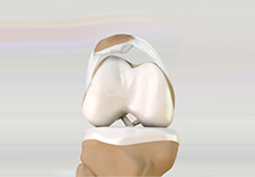
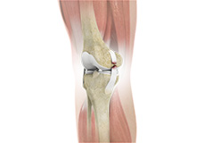
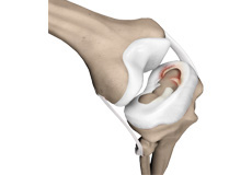
Knee Anatomy
The knee is a complex joint made up of different structures including bones, tendons, ligaments and muscles. They all work together to maintain normal function and provide stability to the knee during movement.
Having a well-functioning healthy knee is essential for our mobility and ability to participate in various activities. Understanding the anatomy of the knee enhances your ability to discuss and choose the right treatment procedure for knee problems with your doctor.
Bones
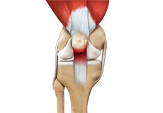
The Knee is a hinge joint made up of two bones, the thigh bone (femur) and the shinbone (tibia). There are two round knobs at the end of the femur called femoral condyles which articulate with the flat surface of the tibia called the tibial plateau. The tibia plateau on the inside of the leg is called the medial tibial plateau, and on the outside of the leg it is called the lateral tibial plateau.
The two femoral condyles form a groove on the front (anterior) side of the knee called the patellofemoral groove. A small bone called the patella sits in this groove and forms the knee cap. It acts as a shield and protects the knee joint from direct trauma.
A fourth bone called the fibula is the other bone of the lower leg. This form a small joint with the tibia. This joint has very little movement and is not considered a part of the main joint of the knee.
Articular Cartilage and Menisci
Movement of the bones causes friction between the articulating surfaces. To reduce this friction, all articulating surfaces involved in movement are covered with a white, shiny, slippery layer called articular cartilage. The articulating surface of the femoral condyles, tibial plateaus and the back of the patella are covered with this cartilage. The cartilage provides a smooth surface that facilitates easy movement.
To further reduce friction between the articulating surfaces of the bones, the knee joint is lined by a synovial membrane which produces a thick clear fluid called synovial fluid. This fluid lubricates and nourishes the cartilage and bones inside the joint capsule.
Within the knee joint between the femur and tibia there are two C shaped cartilaginous structures called menisci. Menisci function to provide stability to the knee by spreading the weight of the upper body across the whole surface of the tibial plateau. The menisci help in load bearing by preventing the weight from concentrating onto a small area, which could damage the articular cartilage. The menisci also act as a cushion between the femur and tibia by absorbing the shock produced by activities such as walking, running and jumping.
Ligaments
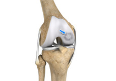
Ligaments are tough bands of tissue that connect one bone to another bone. The ligaments of the knee function to stabilize the knee joint. There are two important groups of ligaments that hold the bones of the knee joint together, collateral ligaments and the cruciate ligament.
Collateral ligaments are present on either side of the knee. They function to prevent the knee from moving too far during side to side motion. The collateral ligament on the inside is called the medial collateral ligament (MCL) and the collateral ligament on the outside is called the lateral collateral ligament (LCL).
Cruciate ligaments- This group of ligaments, present inside the knee joint, control the back and forth motion of the knee. The Cruciate ligament in the front of the knee is called anterior cruciate ligament or ACL and the cruciate ligament in the back of the knee is called posterior cruciate ligament or PCL.
Muscles
Muscles: There are two major muscles, the quadriceps and the hamstrings, which enable movement of the knee joint. The quadriceps muscles are in the front of the thigh. When the quadriceps muscles contract, the knee straightens. The hamstrings are in the back of the thigh. When the hamstring muscles contract, the knee bends.
Tendons
Tendons are structures that attach muscles to the bone. The quadriceps muscles of the knee meet just above the patella and attach to it through a tendon called the quadriceps tendon. The patella further attaches to the tibia through a tendon called the patella tendon. The quadriceps muscle, quadriceps tendon and patellar tendon all work together to straighten the knee. Similarly, the hamstring muscles at the back of the leg are attached to the knee joint with the hamstring tendon.

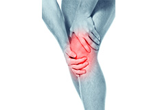
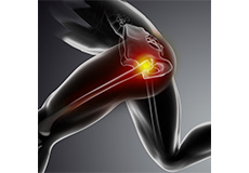
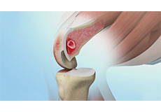
Knee Pain
The knee is one of the largest joints in the body, formed by the lower end of the femur, upper end of the tibia and the patella or knee cap. Several ligaments and muscles attach to the bones of the knee joint to maintain normal motion of the joint. Special cartilaginous tissues known as menisci are placed between the two articular ends of the joint. These act as a cushion between the articular surfaces and absorb the shock during movement.
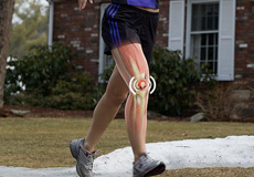
Anterior Knee Pain
Anterior knee pain is a characterized by a chronic pain over the front and center of the knee joint. It is common in athletes, active adolescents (especially girls) and overweight individuals. Anterior knee pain refers to a variety of conditions which include runner's knee or patellar tendinitis and chondromalacia of the patella. There is an inter-individual variation in the duration and presentation of pain.
Runner’s Knee
Runner's knee, also called patellofemoral pain syndrome refers to pain under and around your kneecap. Runner’s knee includes several medical conditions such as anterior knee pain syndrome, patellofemoral malalignment, and chondromalacia patella that cause pain around the front of the knee.
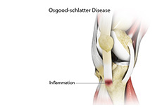
Osgood Schlatter Disease
Osgood-Schlatter disease refers to a condition of an overuse injury that occurs in the knee region of growing children and adolescents. This is caused by inflammation of the tendon located below the knee cap (patellar tendon).
Chondromalacia Patella
The patella, also called the kneecap is a small bone present on the front of your knee joint. The underside of the patella is covered by cartilage that allows smooth gliding of the knee with movement. Overuse or misalignment of the patella can cause wear and tear of the cartilage.
Jumper’s Knee
Jumper’s knee, also known as “patellar tendinitis" is an inflammation of the patellar tendon that connects your kneecap (patella) to your shinbone. This tendon helps in extension of the lower leg.
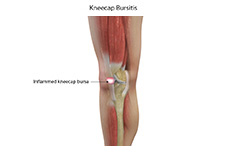
Bursitis
A bursa is a small fluid-filled sac found between soft tissues and bones. It lubricates and acts as a cushion to decrease friction between bones when they move. Bursitis refers to the inflammation and swelling of the bursa. Inflammation of the bursa in front of the kneecap (patella) is known as kneecap bursitis or prepatellar bursitis.
Baker’s Cyst
The knee consists of a fluid called synovial fluid, which reduces friction between the bones of the knee joint while you move your leg. Sometimes this fluid is produced in excess, resulting in its accumulation in the back of your knee. A Baker’s cyst or popliteal cyst is a fluid-filled swelling that develops into a lump behind the knee. This causes stiffness, tightness and pain behind your knee.
Iliotibial Band Syndrome
Iliotibial band syndrome is an overuse injury resulting from the inflammation of iliotibial band. Iliotibial band is a tough group of fibers that begins at the iliac crest of hip and runs along the outside of the thigh, to get attached to the outer side of the shin bone just below the knee joint.
Lateral Patellar Compression Syndrome
Lateral patellar compression syndrome refers to pain under and around your kneecap. It is a common complaint among runners, jumpers, and other athletes such as skiers, cyclists, and soccer players.
Osteochondritis Dissecans
Osteochondritis dissecans is a joint condition in which a piece of cartilage, along with a thin layer of the bone separates from the end of the bone because of inadequate blood supply. The separated fragments are sometimes called “joint mice”. These fragments may be localized, or may detach and fall into the joint space causing pain and joint instability.
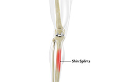
Shin Splints
“Shin splints” is used to describe the pain and inflammation of the tendons, muscles and bone tissue around the tibia or shine bone (a large bone in the lower leg). It occurs because of vigorous physical activity such as exercise or sports. The condition is also referred to as medial tibial stress syndrome (MTSS).

Knee Injury
Pain, swelling and stiffness are the common symptoms of any damage or injury to the knee. If care is not taken during the initial phases of injury, it may lead to joint damage that may end up destroying your knee.
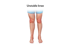
Unstable Knee
The knee joint is one of the largest joints in the body. This highly complex joint has several tissues supporting and stabilizing its movement

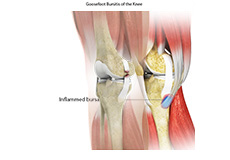
Pes Anserine Bursitis of the Knee
A bursa is a small fluid-filled sac found between soft tissues and bones. It lubricates and acts as a cushion to decrease friction between bones when they move. Bursitis refers to the inflammation and swelling of the bursa. Goosefoot bursitis or pes anserine bursitis is the inflammation of the bursa present between the tendons of the hamstring muscle and the tibia (shinbone) on the inner side of the knee
Knee Sprain
Knee sprain is a common injury that occurs from overstretching of the ligaments that support the knee joint. A knee sprain occurs when the knee ligaments are twisted or turned beyond its normal range causing the ligaments to tear.
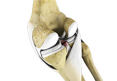
ACL Tears
The anterior cruciate ligament, or ACL, is one of the major ligaments of the knee that is in the middle of the knee and runs from the femur (thigh bone) to the tibia (shin bone). It prevents the tibia from sliding out in front of the femur. Together with posterior cruciate ligament (PCL) it provides rotational stability to the knee.
MCL Tears
The medial collateral ligament (MCL) is the ligament that is located on the inner part of the knee joint. It runs from the femur (thighbone) to the top of the tibia (shinbone) and helps in stabilizing the knee. Medial collateral ligament (MCL) injury can result in a stretch, partial tear, or complete tear of the ligament.
MCL Sprain
The medial collateral ligament (MCL), a band of tissue present on the inside of your knee joint, connects your thigh bone and shin bone (bone of your lower leg). The MCL maintains the integrity of the knee joint and prevents it from bending inward.

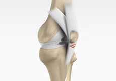
Meniscal Injuries
The knee is one of the most complex and largest joint in the body, and is more susceptible to injury. Meniscal tears are one among the common injuries to the knee joint. It can occur at any age, but are more common in athletes playing contact sports.

Meniscal Tears
Meniscus tear is the commonest knee injury in athletes, especially those involved in contact sports. A suddenly bend or twist in your knee cause the meniscus to tear. This is a traumatic meniscus tear. Elderly people are more prone to degenerative meniscal tears as the cartilage wears out and weakens with age.
Ligament Injuries
The knee is a complex joint which consists of bone, cartilage, ligaments and tendons that make joint movements easy and at the same time more susceptible to various kinds of injuries.
Knee Arthritis
Arthritis is a general term covering numerous conditions where the joint surface or cartilage wears out. The joint surface is covered by a smooth articular surface that allows pain free movement in the joint. This surface can wear out for several reasons; often the definite cause is not known.
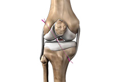
Multiligament Instability
The knee is a complex joint of the body which is vital for movement. The four major ligaments of the knee are anterior cruciate ligament, posterior cruciate ligament, medial collateral ligament and lateral collateral ligament. They play an important role in maintaining the stability of the knee.
Multiligamentous Knee Injury
Ligaments are the fibrous tissue bands connecting the bones in the joint and stabilizing the joint. Knee joint has 2 sets of ligaments–collateral ligaments (medial and collateral ligaments) that connect the bones on outer side of the knee and cruciate ligaments (anterior cruciate ligament and posterior cruciate ligament) those present inside the joint.
Knee Dislocation
Patella (kneecap) is a protective bone attached to the quadriceps muscles of the thigh by quadriceps tendon. Patella attaches with the femur bone and forms a patellofemoral joint. Patella is protected by a ligament which secures the kneecap from gliding out and is called as medial patellofemoral ligament (MPFL).
Patellar Dislocation/Patellofemoral Dislocation
Patella (knee cap) is a protective bone attached to the quadriceps muscles of the thigh by quadriceps tendon. Patella attaches with the femur bone and forms a patellofemoral joint. Patella is protected by a ligament which secures the kneecap from gliding out and is called as medial patellofemoral ligament (MPFL).
PCL Injuries
Posterior cruciate ligament (PCL), one of four major ligaments of the knee are situated at the back of the knee. It connects the thighbone (femur) to the shinbone (tibia). The PCL limits the backward motion of the shinbone.
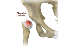
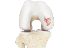
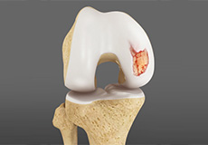
Chondral (Articular Cartilage Defects)
Articular or hyaline cartilage is the tissue lining the surface of the two bones in the knee joint. Cartilage helps the bones move smoothly against each other and can withstand the weight of the body during activities such as running and jumping.
Patellar Instability
Patellar (knee cap) instability results from one or more dislocations or partial dislocations (subluxations). Patella is the small piece of bone in front of the knee that slides up and down the femoral groove (groove in the femur bone) during bending and stretching movements.
Patellofemoral Instability
The knee can be divided into three compartments: patellofemoral, medial and lateral compartment. The patellofemoral compartment is the compartment in the front of the knee between the knee cap and thigh bone. The medial compartment is the area on the inside portion of the knee, and the lateral compartment is the area on the outside portion of the knee joint.
Patella Fracture
The knee cap or patella is the largest sesamoid bone in the body and one of the components of the knee joint, present at the front of the knee. The undersurface of the kneecap and the lower end of the femur are coated with articular cartilage, which helps in smooth movement of the knee joint. The knee cap protects the knee and provides attachment to various muscle groups of the thigh and leg. Fracture of knee cap is rare and is more common in adult males.
Recurrent Patella Dislocation
The patella (knee cap) is a small bone that shields your knee joint. It is found in front of your knee, in a groove called the trochlear groove that sits at the junction of the femur (thighbone) and tibia (shinbone). Articular cartilage present below the patella and end of the femur cushion and help the bones glide smoothly over each other when the legs move. This joint is stabilized and supported by a network of soft tissues.
Quadriceps Tendon Rupture
Quadriceps tendon is a thick tissue located at the top of the kneecap. The quadriceps tendon works together with the quadriceps muscles to allow us to straighten our leg. The quadriceps muscles are the muscles located in front of the thigh.

Patella Tendon Rupture
Patella tendon rupture is the rupture of the tendon that connects the patella (knee cap) to the top portion of the tibia (shin bone). The patellar tendon works together with the quadriceps muscle and the quadriceps tendon to allow your knee to straighten out.
Lateral Meniscus Syndrome
The knee joint is formed by the union of two bones, namely the femur (thigh bone) and the tibia (lower leg bone). At the junction of these two bones is a cartilage called the meniscus, which acts as a shock absorber. There are two menisci – the lateral and medial menisci.
Medial Meniscus Syndrome
Of the menisci within the knee, it is the medial that is more easily injured. Differences in the anatomical attachments of the medial meniscus compared to the lateral mean that the medial meniscus becomes distorted during combined flexion and rotation movements in a manner not experienced on the lateral side.
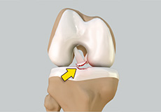
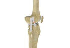
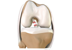
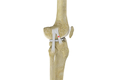
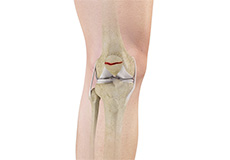
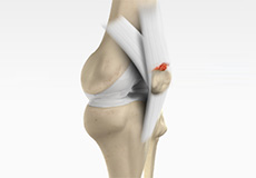
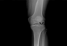
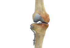
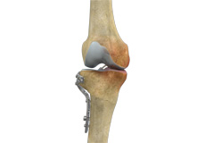
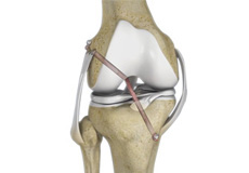
Tibial Eminence Spine Avulsions
Tibial eminence spine avulsion fracture is avulsion (tear away) of the tibial eminence (an extension on the bone for attachment of muscles) which most commonly involves the anterior cruciate ligament (ACL) insertion site.
Osteonecrosis of the Knee
Osteonecrosis is a condition in which death of a section of bone occurs because of lack of blood supply to it. It is one of the most common causes of knee pain in older women. Women over the age of 60 years of age are commonly affected, three times more often than men.
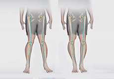
Knee Angular Deformities
Angular deformities of the knee are common during childhood and usually are variations in the normal growth pattern. Angular deformity of the knee is a part of normal growth and development during early childhood.
Non-surgical Treatments

Pharmacological
The knee is a complex joint which consists of bone, cartilage, ligaments and tendons that make joint movements easy and at the same time more susceptible to various kinds of injuries. Knee problems may arise if any of these structures get injured by overuse or suddenly during sports activities. Injuries to the knee can be caused by degenerative disease such as arthritis, traumatic injuries and sports injuries.
Platelet Rich Plasma Injection (PRP)
Our blood consists of a liquid component known as plasma. It also consists of three main solid components which include the red blood cells (RBCs), white blood cells (WBCs), and platelets. Platelets play an important role in forming blood clots. They also consist of special proteins, known as growth factors, which help with our body’s healing process. Platelet-rich plasma or PRP is a high concentration of platelets and plasma.
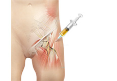
Stem Cell Treatments for the Knee
Stem cell therapy is a form of regenerative medicine that utilizes the body’s natural healing mechanism to treat various conditions.
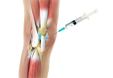
Viscosupplementation
Viscosupplementation refers to the injection of a hyaluronan preparation into the joint. Hyaluronan is a natural substance present in the joint fluid that assists in lubrication. It allows smooth movement of the cartilage covered articulating surfaces of the joint.
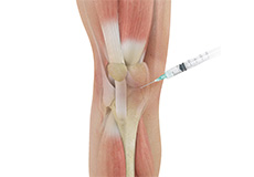
Cortisone Injection
Cortisone is a corticosteroid released by the adrenal gland in response to stress and is a potent anti-inflammatory agent. Artificial preparations containing cortisone are injected directly into the affected joint to relieve pain and reduce inflammation.
Physical Therapy
Physiotherapy or physical therapy is an exercise program that helps you to improve movement, relieve pain, encourage blood flow for faster healing, and restore your physical function and fitness level. The main aim of physical therapy is to make your daily activities such as walking, getting in and out of bed, or climbing stairs easier. It can be prescribed as an individual treatment program or combined with other treatments.
Surgical Treatment
Knee Osteotomy
Knee Osteotomy is a surgical procedure in which the upper shinbone (tibia) or lower thighbone (femur) is cut and realigned. It is usually performed in arthritic conditions affecting only one side of your knee and the aim is to take pressure off the damaged area and shift it to the other side of your knee with healthy cartilage.
High Tibial Osteotomy
High tibial osteotomy is a surgical procedure performed to relieve pressure on the damaged site of an arthritic knee joint. It is usually performed in arthritic conditions affecting only one side of your knee and the aim is to take pressure off the damaged area and shift it to the other side of your knee with healthy cartilage.
Tibial Tubercle Osteotomy
Tibial tubercle osteotomy is a surgical procedure which is performed along with other procedures to treat patellar instability, patellofemoral pain, and osteoarthritis. This is a quite safe procedure and provides excellent access and surgical exposure during a difficult primary or revision total knee arthroplasty.
Medial Patellofemoral Ligament Reconstruction
Medial patellofemoral ligament reconstruction is a surgical procedure indicated in patients with more severe patellar instability. Medial patellofemoral ligament is a band of tissue that extends from the femoral medial epicondyle to the superior aspect of the patella.
Distal Realignment Procedures
Distal realignment procedures, also known as TTT or tibial tubercle transfer procedures are performed to reposition the kneecap by realigning the tendon under the kneecap to the underlying tibial tubercle. Tibia tubercle is the bony lump on the tibia (bone in the lower leg) below the knee cap.
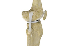
Arthroscopic Reconstruction of the Knee for Ligament Injuries
The knee is the most complex joint in the body and is formed by the articulation between the thigh bone (femur) and the shinbone (tibia). A knee cap is present over the front of the joint to provide extra protection. These bones are held together by four strong rope like structures called ligaments. Two collateral ligaments are present on either side of the knee and control the sideway movements of the knee.
PCL Reconstruction
Posterior cruciate ligament (PCL), one of four major ligaments of the knee are situated at the back of the knee. It connects the thighbone (femur) to the shinbone (tibia). The PCL limits the backward motion of the shinbone.
LCL Reconstruction
Lateral collateral ligament (LCL) is a thin set of tissues present on the outer side of the knee, connecting the thighbone (femur) to the fibula (side bone of lower leg). It provides stability as well as limits the sidewise rotation of the knee. Tear or injury of LCL may cause instability of the knee that can be either reconstructed or repaired to regain the strength and movement of the knee.

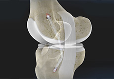
ACL Reconstruction
The anterior cruciate ligament is one of the major stabilizing ligaments in the knee. It is a strong rope like structure located in the center of the knee running from the femur to the tibia. When this ligament tears unfortunately, it does not heal and often leads to the feeling of instability in the knee.
Cartilage Restoration
Patellar Tendon Repair
Patella tendon rupture is the rupture of the tendon that connects the patella (knee cap) to the top portion of the tibia (shin bone). The patellar tendon works together with the quadriceps muscle and the quadriceps tendon to allow your knee to straighten out.
Knee Ligament Reconstruction
The knee is the most complex joint in the body and is formed by the articulation between the thigh bone (femur) and the shinbone (tibia). A knee cap is present over the front of the joint to provide extra protection. These bones are held together by four strong rope like structures called ligaments. Two collateral ligaments are present on either side of the knee and control the sideway movements of the knee.
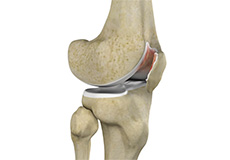
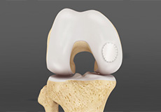
Cartilage Replacement
Cartilage replacement is a surgical procedure performed to replace the worn-out cartilage with the new cartilage. It is usually performed to treat patients with small areas of cartilage damage usually caused by sports or traumatic injuries. It is not indicated for those patients who have advanced arthritis of knee.
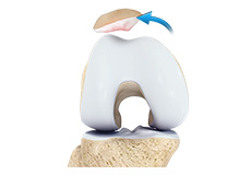
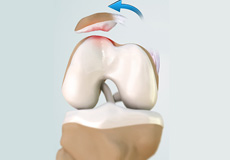
Cartilage Repair and Transplantation
Articular Cartilage is the white tissue lining the end of bones where these bones connect to form joints. Cartilage acts as cushioning material and helps in smooth gliding of bones during movement. An injury to the joint may damage this cartilage which cannot repair on its own.
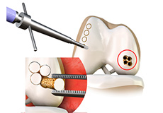
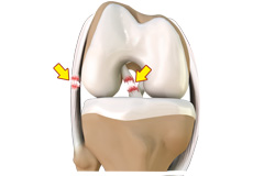
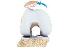
OATS
OATS is “osteochondral autograft transfer system”. It is one of the two types of cartilage transfer procedures and the other procedure is “Mosaicplasty”. Cartilage transfer procedures involve moving healthy cartilage from a non-weight bearing area of the knee to a damaged area of the cartilage in the knee.
Cartilage Restoration
Articular cartilage is the smooth, shiny, white tissue covering the ends of bones those form a joint. Articular cartilage reduces friction when bones glide over each other, making the movements smooth and painless. It also acts as a shock-absorber to help prevent traumatic injuries to the bones. When cartilage is damaged it can cause painful movements and limited joint mobility and eventually progress to osteoarthritis.
Others
Autologus Chondrocyte Implantation
Autologous chondrocyte implantation (ACI) is a procedure to treat the articular cartilage defects of the knee. This procedure is effective for treating small areas of cartilage damage that causes pain and swelling and restricts the range of motion. Autologous chondrocyte implantation is not indicated for those patients who have advanced arthritis of knee.
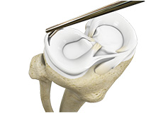
Partial Meniscectomy
Partial meniscectomy is a surgical procedure to remove the torn portion of the meniscus from the knee joint. Meniscus is the C-shaped cartilage located in the knee that lubricates the knee joint, acts as shock-absorber, and controls the flexion and extension of joint. Meniscal tears can occur at any age, but are more common in athletes playing contact sports.
Meniscal Surgery
Meniscus tear is the commonest knee injury in athletes, especially those involved in contact sports. A suddenly bend or twist in your knee cause the meniscus to tear. This is a traumatic meniscus tear. Elderly people are more prone to degenerative meniscal tears as the cartilage wears out and weakens with age.
Subchondroplasty
Knee osteoarthritis (OA) is a common form of arthritis that causes joint pain and stiffness. It is a progressive disease in which the joint cartilage gradually wears away and may lead to disability.
Meniscal Tear Repair
Meniscus tear is the commonest knee injury in athletes, especially those involved in contact sports. A suddenly bend or twist in your knee cause the meniscus to tear. This is a traumatic meniscus tear. Elderly people are more prone to degenerative meniscal tears as the cartilage wears out and weakens with age.