Fractures & Trauma
What is a Fracture?
A bone fracture is a medical condition in which a bone is cracked or broken. It is a break in the continuity of the bone. While many fractures are the result of high force impact or stress, bone fracture can also occur because of certain medical conditions that weaken the bones, such as osteoporosis.
The word “Fracture” implies to broken bone. A bone may get fractured completely or partially and it is caused commonly from trauma due to fall, motor vehicle accident or sports. Thinning of the bone due to osteoporosis in the elderly can cause the bone to break easily. Overuse injuries are common cause of stress fractures in athletes.
Types of fractures include:
- Simple fractures in which the fractured pieces of bone are well aligned and stable.
- Unstable fractures are those in which fragments of the broken bone are misaligned and displaced.
- Open (compound) fractures are severe fractures in which the broken bones cut through the skin. This type of fracture is more prone to infection and requires immediate medical attention.
- Greenstick fractures: This is a unique fracture in children that involves bending of one side of the bone without any break in the bone.
Fracture Healing
Our body reacts to a fracture by protecting the injured area with a blood clot and callus or fibrous tissue. Bone cells begin forming on the either side of the fracture line. These cells grow towards each other and thus close the fracture.
Medical Therapy
The objective of early fracture management is to control bleeding, prevent ischemic injury (bone death) and to remove sources of infection such as foreign bodies and dead tissues. The next step in fracture management is the reduction of the fracture and its maintenance. It is important to ensure that the involved part of the body returns to its function after fracture heals. To achieve this, maintenance of fracture reduction with immobilization technique is done by either non-operative or surgical method.
Non-operative (closed) therapycomprises of casting and traction (skin and skeletal traction).
- Casting closed reduction is done for any fracture that is displaced, shortened, or angulated. Splints and casts made up of fiberglass or plaster of Paris material are used to immobilize the limb.
- Traction Traction method is used for the management of fractures and dislocations that cannot be treated by casting. There are two methods of traction namely, skin traction and skeletal traction.
Skin traction involves attachment of traction tapes to the skin of the limb segment below the fracture. In skeletal traction, a pin is inserted through the bone distal to the fracture. Weights will be applied to this pin, and the patient is placed in an apparatus that facilitates traction. This method is most commonly used for fractures of the thighbone.
Surgical Therapy
- Open Reduction and Internal Fixation (ORIF)
This is a surgical procedure in which the fracture site is adequately exposed and reduction of fracture is done. Internal fixation is done with devices such as Kirschner wires, plates and screws, and intramedullary nails. - External fixation
External fixation is a procedure in which the fracture stabilization is done at a distance from the site of fracture. It helps to maintain bone length and alignment without casting.
External fixation is performed in the following conditions:
- Open fractures with soft-tissue involvement
- Burns and soft tissue injuries
- Pelvic fractures
- Comminuted and unstable fractures
- Fractures having bony deficits
- Limb-lengthening procedures
- Fractures with infection or non-union
Rehabilitation
Fractures may take several weeks to months to heal completely. You should limit your activities even after the removal of cast or brace so that the bone become solid enough to bear the stress. Rehabilitation program involves exercises and gradual increase in activity levels until the process of healing is complete.
Knee

Fractures of the Proximal Tibia
The tibia or shin bone is a major bone of the leg which connects the knee to the ankle. A tibial fracture is a break in the continuity of the shin bone (tibia).
Fractures of proximal tibia: A proximal tibial fracture is a break in the upper part of the shin bone or tibia.
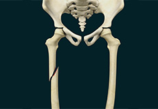
Pediatric Thighbone (Femur) Fracture
The femur or thighbone is the largest and strongest bone in the human body. Pediatric thighbone fractures can occur when your child falls hard on the ground and gets hit during sports, automobile accidents, and child abuse. In a thighbone fracture, the broken bones may be aligned or displaced. The fracture can either be closed (with skin intact) or open (with the bone piercing out through the skin).
Thighbone (Femur) Fracture
The femur or thighbone is the longest and strongest bone in the body, connecting the hip to the knee. A femur fracture is a break in the femur. The distal femur is the lower part of the thigh bone which flares out like an upside-down funnel and its lower end is covered by a smooth, slippery articular cartilage that protects and cushions the bone during movement. Fracture of the distal femur may involve the cartilaginous surface of the knee as well and result in arthritis.
Shoulder, Elbow
Broken Collarbone
The clavicle or the collarbone is the bone that connects your sternum or breastbone to your shoulder. Clavicle fracture, also called broken collarbone is a very common sports injury seen in people who are involved in contact sports such as football and martial arts as well as impact sports such as motor racing.
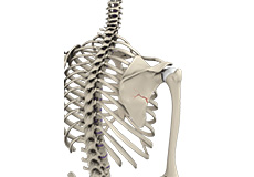
Fracture of the Shoulder Blade (Scapula)
The scapula (shoulder blade) is a flat, triangular bone providing attachment to the muscles of the back, neck, chest and arm. The scapula has a body, neck and spine portion.
Scapular fractures are uncommon but do occur and require a large amount of force to fracture.
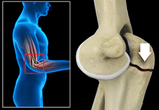
Olecranon (Elbow) Fractures
Three bones, humerus, radius and ulna make up the elbow joint. The bones are held together by ligaments thus providing stability to the joint. Muscles and tendons around the bones coordinate the movements and help in performing various activities. Elbow fractures may occur from trauma resulting from a variety of reasons, some of them being a fall on an outstretched arm, a direct blow to the elbow, or an abnormal twist to the joint beyond its functional limit.
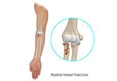
Radial Head Fractures
The elbow is a junction between the forearm and the upper arm. The elbow joint is made up of 3 bones namely the humerus bone in the upper arm which joins with the radius and ulna bones in the forearm. The elbow joint is essential for the movement of your arms and to perform daily activities. The head of the radius bone is cup-shaped and corresponds to the spherical surface of the humerus. The injury in the head of the radius causes impairment in the function of the elbow.
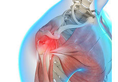
Shoulder Trauma
Shoulder injuries most commonly occur in athletes participating in sports such as swimming, tennis, pitching, and weightlifting. The injuries are caused due to the over usage or repetitive motion of the arms.